Ventricular pause 의 4가지 원인
- 1. Blocked APC (Non conducted APC)
- 2. 2nd degree AV block or High degree AV Block
- 3. SA Block
- 4. Sinus arrest
QRS 앞에 P wave 가 존재할 경우 가장 긴 T-P segment 사이를 살펴본다.
* P wave 가 없다면
- (1) Sinus node dysfunction – P wave 를 만들지 못했다.
- (2) SA exit block – P wave 는 만들었지만 전도가 안되었다. (배수의 관계)
* P wave 가 있다면
- (1) Non conducted APC
- (2) VPC with retrograde concealed block
- (3) 2nd degree 이상의 AV block
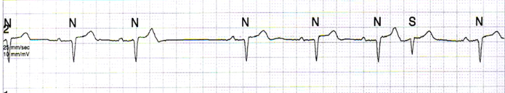
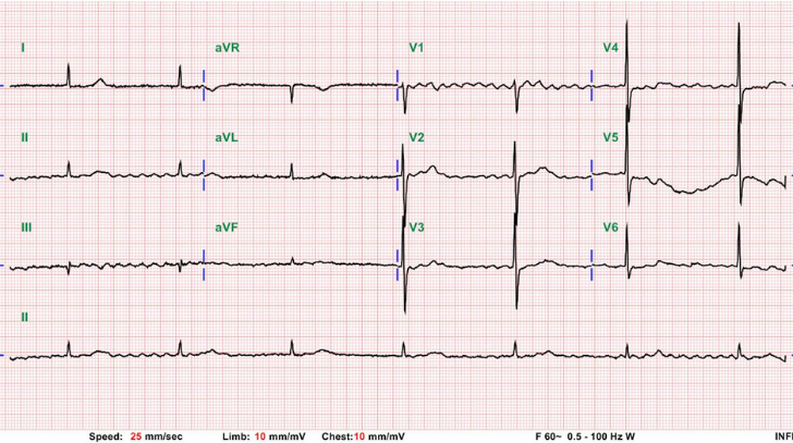
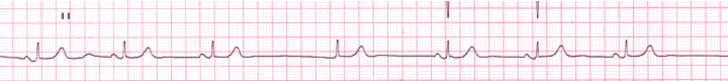
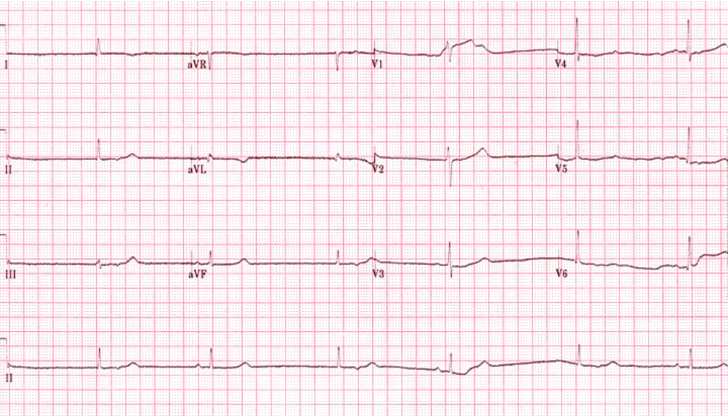
Non conducted APC
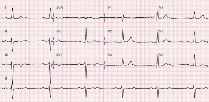
2nd degree AV block Mobitz type 1
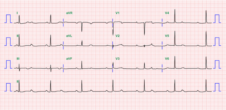
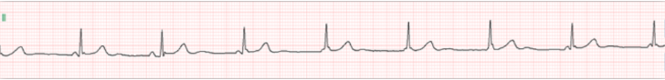
2:1 AV block
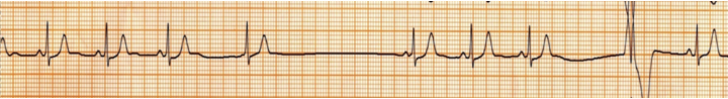
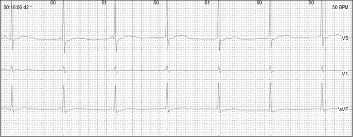
Complete AV Block
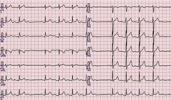
Complete AV block with permanent AF
QRS 앞에 P wave 가 존재하지 않을 경우
- 1. Junctional escape rhythm
- 2. Ventricular escape rhythm
- 3. Ectopic atrial rhythm
- 4. Isorhythmic AV dissociation
* Sinus node dysfunction with junctional escape beat
* Sinus node dysfunction with Ventricular escape beat
* Junctional rhythm with ectopic atrial beat
* Isorhythmic AV dissociation
Sinus node dysfunction with junctional escape beat
(Isorhythmic) AV Dissociation
Two separate pacemakers in A and V (Drug, Ischemia, Local irritation)
–> 쉽게말해 First pacemaker 가 60~100 회는 뛰어주어야 하는데, 40~60 회 밖에 뛰지를 못하니, Second pacemaker 가 작동하기 시작하였고, First pacemaker 가 전달되어 심실 수축을 이끌었지만, 이 후에 first pacemaker 나오는 것이 늦어 second pacemaker 가 나오면서 서로 경쟁하는 듯한 모습을 보이는 현상.
–> AV junctional rhythm d/t Severe sinus bradycardia
–> 증상이 없다면 치료 필요 없다.
* AV Dissociation ⊃ Complete AV block
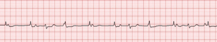
Interference AV dissociation
first pacemaker 는 60~100 회로 정상적으로 뛰어주고 있는데, Second pacemaker 가 항진이 되어 40~60 회가 아닌 60~100 회를 뛰어 마치 경쟁하는 듯한 모습을 보이는 현상
대표적인 예로 Reperfusion arrhythmia 의 예 (AIVR) 가 있다.
–> Enhanced Lower (junctional or Ventricular) pacemaker
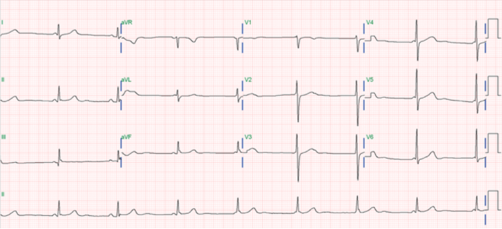
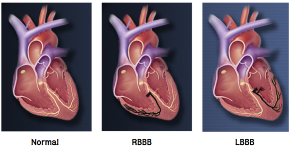
BBB (Bundle branch block)
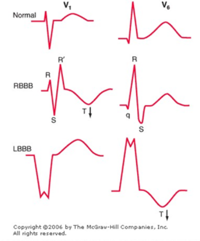
–> V1 과 V6 은 서로 mirror image (Vector 가 반대)
각 차단을 동반한 Bradycardia 시, QRS axis, PR 간격, T wave 의 형태를 확인.
- 1. QRS Axis : II, III, aVF lead 를 보고 superior axis 는 아닌지
- 2. PR interval : 200 ms 이상 증가하였는지
- 3. T-wave : Superimposed P wave 는 아닌지.
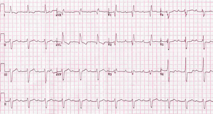
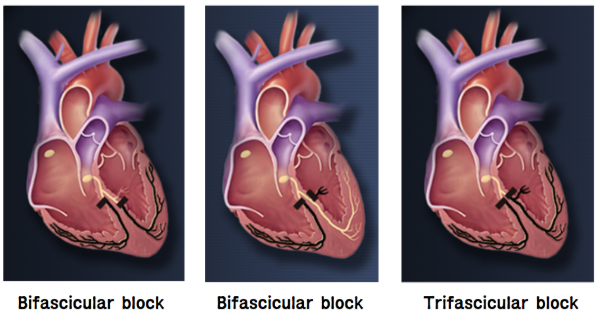
- 1) 1st degree AV block 이 있다.
- 2) RBBB 가 있다.
- 3) II, III, AVF 에서 QRS 가 반대인 Superior axis 이다. –> LAFB
–> Tri-Fascular Block **
심박동기의 적응증
SSS
– Symptomatic sinus bradycardia, sinus pause, chronotropic incompetence
AV block
- Symptomatic (including heart failure or ventricular arrhythmia due to AV block) 3rd degree and 2nd degree AV block
- Symptom free 3rd degree and advanced 2nd degree AV block :
- >3sec (SR) or >5sec (AF) pause, < 40bpm, escape rhythm below AV node
- 3rd degree and 2nd degree AV block during exercise
MRI 촬영시 심박동기
– MRI 가능한 device 라도 old lead, 다른 device 유무 확인이 필요함
- Threshold 가 2.0V 이상은 불가능
- Implantation 후 6주 이후에 촬영 가능
- 촬영 전후 mode change 및 programming 필요