inspiration 촬영 vs expiration 촬영
expiration 했을 시
1. heart 와 vessel 이 커져보인다.
2. lung 이 whiter 해 보인다.
3. diaphragm 이 올라간다.
expiration film 의 유용성
1. to detect focal air trapping from asymmetrical emphysema
2. to detect partial bronchial obstruction (air trappping)
– air trapping 이 있는 상태에서 expiration film 촬영 시, 정상 폐는 whiter 하게 되고 병변이 있는 쪽 폐는 계속 검게 남아있다. (unchanged)
3. accentuate a small pneumothorax
– expiration 시 deflation 된 폐는 whiter 해지고, intrapleural air 는 black 영역으로 커져 구별하기 쉬워짐.
four basic tissue density
– air / fat / water (soft tissue) / metal (bone)
– air = -1000 HU
– lung = -800 HU
– fat = -80~120 HU
– fluid = 0 HU (pure water = 0 HU)
– muscle = +40 HU (soft tissue densitiy)
– bone >= +350 HU
Viewing sequence
– ATMLL (are there many lung lesion ?)
– Abdomen / Thorax / Mediastinum / Lung – unilateral / Lungs – bilateral
Lung parenchyma
– air sacs (alveoli) + supporting structures (interstitium)
– alveoli 는 모여 acini 형태로 terminal airway 주위에 배치되어 있음.
– 몇개의 acini 가 모여 secondary pulmonary lobule 을 형성
=> basic unit of lung function and gross morphology
Interstitium
– supporting the alveoli — supporting framework
==> vessels, lymphatics, bronchi, connective tissue
– CXR 에서는 pulmonary a. and v. 만 눈에 보임. 사실상 interstitium 에서. (interstitial marking)
==> branch 끝으로 갈 수록 tapper 해 가다가 말단 1/3 에서는 거의 보이지 않음. (매우 작음)
– interstitium 에 disease 가 생기면, interlobular septa 나 small vessels 를 둘러싸는 interstitial tissue 가 thicken해짐. 그러면 말단부로 갈 수록 안보여야하는데, 잘 보이게됨.
==> interstitial thickening
Alveoli
– fluid 나 tissue 로 air sacs 이 채워지면 radiodense 해짐.
==> alveolar consolidation, homogeneously white (not aerated)
– interstitial marking 은 잘 안보임.
3가지 accessory fissures
1. azygos fissure — azygos lobe
2. inferior accessory fissure
3. superior accessory fissure
silhouette sign
– RUL — ascending aorta, Rt. tracheal lung interface
– RML — Rt. heart
– RLL — Rt. diaphragm
– LUL — Lt. atrium, Aortic arch, Ant./mid. mediastinum
– lingula — Lt. heart
– LLL — Lt. diaphragm
air bronchogram sign
– visualization of the bronchi (Air bronchogram) indicates a pulmonary lesion
(not pleural lesion / not mediatinal lesion)
==> bronchi contain air and the adjacent lung is consolidated
* 단 Air bronchogram 이 안나타나는 조건이 있다!
(1) secretion 이나 tumor 가 bronchi 를 막았을 경우
(2) patchy peripheral lung consolidation
(3) interstitial disease
(4) hyperinflated condition — asthma 등
* cardiac shadow 뒤에 air bronchogram 이 보인다면, LLL consolidation 의 증거가 된다.
Lung and lobar Collapse
RML/Lingular collapse 는 PA 로 찍으면 subtle 할 수 있다. lateral 이 더 잘 보인다.
Bronchus intermedium 는 RML, RLL 동시에 연결되어 있어 RML, RLL 동시에 collapse 잘 된다.
(LUL, lingula 도 마찬가지)
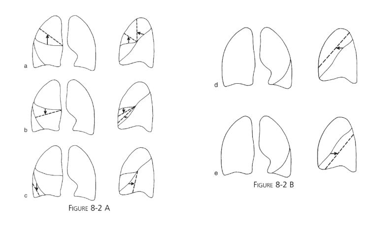
The best and most frequent sign of lobar collapse ==> fissure movement
Two less frequent signs of lung collapse ==> crowded bronchi and vessels / moving marker structures
hillum 이 내려오는 것은 lower lobe collapse 를 시사
hillum 이 올라가는 것은 upper lobe collapse 를 시사
Middle lobe 이나 lingula collapse 는 hillum 변화 없음.
(* Left hilum 은 정상적으로 Right hilum 보다 살짝 위에 있음. — 97%)
e.g. >
LLL collapse 시
1) Lt. hillum 위치가 내려와 Rt. 와 비슷하게 된다.
2) Lt. Diaphragm 이 올라간다.
3) Left lung 이 more radiolucent 해진다.
4) 심장 뒷쪽으로 air bronchogram 이 생기면서 bronchi 가 모여있다.
Collapse 의 physiologic mechanisms
(1) obstructive (resorptive) atelectasis
– airway 는 어느 한 곳이 막히면 막힌 곳의 distal 부위의 air 는 훕수된다.
– Central obstruction : main, lobar, segmental bronchus
; no air bronchogram sign
==> intrinsic (endobronchial tumor) or extrinsic (tumor around bronchus)
– Peripheral obstruction : multiple small mucous plugs, blood clots, in small bronchi
; air bronchograms are present
(2) passive (relaxation) atelectasis
– e.g. hydrothorax 나 pneumothorax 때문에 눌려서.
(3) cicatricial (scarring) atelectasis
– local : TBc, radiation 등에 의한 pulmonary fibrosis 때문
– general : silicosis, sarcoidosis
(4) adhesive atelectasis (surfactant 의 감소)
(5) hypoventilation atelectasis (CNS 문제 또는 Pain 문제)
– 주로 전신마취 후 lung base 에 자주 생긴다.
Plate or bandlike atelectasis
: atelectasis 가 segmental level 이나 random small areas of the lung parenchyma 에 생기는 경우