
Light microscopy
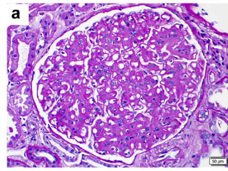
FGN 의 광학현미경 소견에서 가장 흔한 사구체 손상의 패턴은 mesangial GN 으로, 전체 케이스의 21~78% 경우에 해당되었다. 이는 deposits and sclerosis accompanied by variable degrees of mesangial hypercellularity 로 인해 발생하는 mesangial expansion 을 특징으로 한다. (그림 a)
두번째로 흔한 패턴은 MPGN 이고 전체 케이스의 11%~56% 에 해당한다. MPGN 은 segmental or global glomerular basementmembrane double contouring and cellular interposition 이 특징이다. 이러한 패턴은 항상 mesangial sclerosis, deposits, and hypercellularity 와 관련되어 있으며, 따라서 advanced stage of disease 를 나타낸다. 시간에 따라서 mesangial GN 에서 MPGN 으로 transformation 한다는 보고는 이러한 내용을 뒷받침한다.
드문 경우에는 membranous-like GN 으로 나타나는 경우가 있는데, 전체 케이스 중 0%–19% 를 차지하였다. 이 패턴은 특징적으로 global glomerular basement membrane thickening and subepithelial infiltration of fibrils with spike formation 을 보였다.
또 다른 드문 케이스로 endocapillary proliferative GN 이 있고, 이는 전체 케이스 중 0%~15% 를 차지했다. 특징으로 endocapillary hypercellularity and leukocyte infiltration 이 있는데, 이는 peripheral capillaries 의 폐색으로 발생한다. 상기 두 가지 드문 패턴의 경우에도 역시 보통은 mesangial deposits, sclerosis, and hypercellularity 와 연관이 있었다.
Focal cellular or fibrous crescents 의 경우 전체 케이스의 17% ~ 50% 에서 보였다. 하지만 50% 이상의 사구체가 침범당하는 diffuse crescentic pattern 은 드물었고 단지 4~6% 정도밖에 되지 않았다.
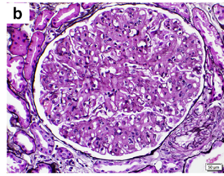
FGN 에서 mesangial deposits 은 (glassy) 유리같고, 다양한 주기의 PAS positive 양상 (variably periodic acid–Schiff positive) 이었으며, nonargyrophilic (은친화가 아닌) 이었다. (그림 a, b)
비록 FGN 의 전통적인 정의에 기초한다면 deposits 은 반드시 Congo red–negative 여야 하지만, a 최근 Congo red positive FGN (defined by polyclonal IgG and DNAJB9 positivity)의 18 개 케이스를 분석한 임상적, 병리적 연구가 발표되었으며, 이는 amyloidosis 와 혼동될 수 있다.
Immunofluorescence microscopy
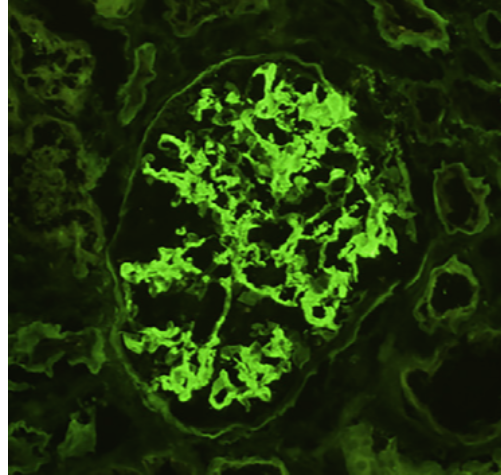
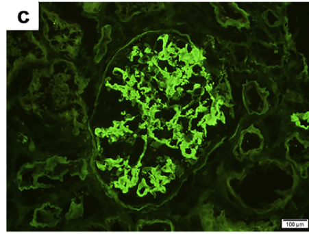
대부분의 FGN 의 경우 polyclonal IgG 과 C3 의 면역형광염색에 대해 intense mesangial and glomerular capillary wall 부분이 양성을 보여준다. 하지만 IgA, IgM, and C1q 염색은 glomerular staining 이 흔하지 않고, intense 하지도 않다. (그림 c)
glomerular deposits 의 텍스쳐는 보통 smudgy (지저분, 얼룩덜룩) 하다. 적은 수의 케이스에서는 anti–glomerular basement membrane GN 과 비슷한 양상의 pseudolinear staining of the glomerular basement membrane 을 보여주기도 한다.
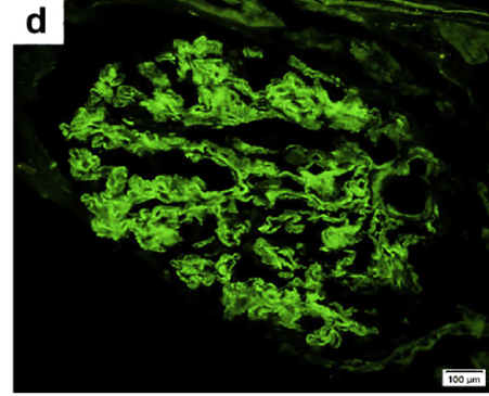
FGN 의 대부분의 케이스에서 IgG deposits 은 polytypic 했다. 3%–15% 정도의 적은 경우에는 monotypic deposits 을 보였다. IgG subclass analysis 에서 대부분의 FGN 에서는 IgG4 에 염색이 되었다. (그림 d) 반면 IgG1 이 우세하거나 혼재되어 염색되는 경우가 드물게 있었다.
뚜렷하게 light chain 으로만 구성된 deposit 이 침작된 몇몇 FGN 사례 (특히 이들은 lambda 로만 구성) 의 frozen tissue 의 경우 protease-digested, paraffin-embedded tissue 에서 kappa 와 lambda 모두가 면역형광 염색이 되는 것을 보여주었다. 이러한 케이스들은 실제로 monoclonal gammopathy 의 임상적 양상은 아니었다.
따라서, monoclonal FGN 은 오로지 paraffin immunofluorescence 에 의해 light chain restriction 과 immunofluorescence staining for IgG subclasses 에 의해 IgG subtype restriction 이 컨펌이 된 이 후에나 진단이 가능해진다.
Multiple myeloma 나 high-grade lymphoma 가 없는 상태에서 Monoclonal FGN 은 monoclonal gammopathy of renal significance lesion 이다.
비록 사구체 외 deposits 은 초기연구에서는 거의 보고가 없었으나, 최근 연구에서는 extraglomerular IgG staining 이 30% ~ 49% 의 케이스에서 나타났다는 보고가 있으며, 가장 흔하게 tubular basement membranes 에 나타났고, 드물게는 few peritubular capillaries or arterioles 에 나타났다.
Electron microscopy
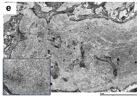
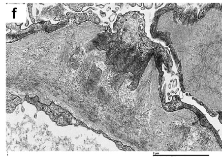
Ultrastructurally, FGN 은 glomerular deposition of randomly oriented straight fibrils that resemble amyloid fibrils but are larger (12–24 nm vs. 8–15 nm) 의 특징이 있다. (그림 e, f)
그러나 여기에는 FGN 과 amyloidosis fibrils 두께 간에 overlap 이 있다. 따라서 fibril diameter 는 구분하는 절대적인 기준이 되지 못한다.
위에서 언급했던대로 congophilic properties of deposits, as Congo red–positivity 특히 그 강도가 약할 때는 그것만 가지고 amyloid 진단을 해서는 안된다.
FGN fibrils 은 거의 항상 mesangium 을 침범하여 (그림 e) 거기서 masangial matrix 와 서로 섞인다. 대부분의 경우에서 fibrils 은 또한 lamina densa of the glomerular basement membrane 에 침윤한다.
때로는 glomerular basement membrane 바깥쪽에 extensive localization of fibrils 이 있을 수 있는데, 이렇게 되면 spike-like projections into the urinary space 가 형성된다. (그림 f)
FGN 에서 Extraglomerular fibrils 는 드물게 보고가 되고 있다. Extraglomerular structures 에 침범되는 경우가 매우 적기도 하고, ultrastructurally 분석된 조직이 별로 없기 때문이다. 저자가 속한 기관의 경우 extraglomerular FGN fibrils (involving rare tubular basement membranes and less commonly peritubular capillaries, arterioles, or interstitium) 이 확인되는 경우는 extensive ultrastructural search 한 전체 케이스의 19% 정도 되었다.
Splenic involvement 외에 other organ involvement (such as lung, heart, and liver) 의 보고는 드물다. 그렇지만 이러한 보고들은 조직내의 FGN fibrils 과 matrix fibrils 의 구분이 어렵기 때문에 확실이 증명되지는 않았다.
- 2020/02/03 – Fibrillary glomerulonephritis, 원섬유성 사구체신염 (1)
- 2020/02/03 – Fibrillary glomerulonephritis, 원섬유성 사구체신염 (2) : Demographics and associated conditions
- 2020/02/03 – Fibrillary glomerulonephritis, 원섬유성 사구체신염 (3) : Clinical renal characteristics and serologies, Outcomes, Treatment
- 2020/02/03 – Fibrillary glomerulonephritis, 원섬유성 사구체신염 (4) : Pathologic characteristics
- 2020/02/03 – Fibrillary glomerulonephritis, 원섬유성 사구체신염 (5) : Identification of DNAJB9 as an excellent diagnostic marker for FGN
- 2020/02/04 – Fibrillary glomerulonephritis, 원섬유성 사구체신염 (6) : A potential pathogenetic role of DNAJB9 in FGN