유방초음파검사
우리나라 여성의 유방은 서양 여성에 비해 크기가 작고 치밀형 유방이 많기에 초음파검사가 용이합니다.
유방초음파 검사는 방사선 조사의 위험성이 없어
30세 이하의 젊은 여성, 임신 또는 수유 중인 여성에서 유방검사가 필요한 경우 일차적 검사 방법입니다.
유방초음파 검사는 일차적으로 만져지는 종괴 또는 유방촬영술에서 발견된 종괴가
고형 종괴와 낭성 종괴의 감별, 고형 종괴 중 양성과 악성의 감별진단에 유용합니다.
다만, 유방초음파검사 단독으로 유방암 검진을 하는 것은
미세석회화나 작은 크기의 유방암을 발견할 수 없으므로 부적절합니다.
작은 고형 병변, 특히 주위 지방에 둘러싸여 있는 병변은 전문의사가 아니라면 초음파로 발견하기 어렵습니다.
또한 높은 위양성율 또한 유방초음파검사의 단점입니다.
그러므로 유방초음파 검사는 유방촬영술의 이상 또는 만져지는 부위에 대한 문제해결 차원에서 시행되어야 하겠습니다.
정상유방의 초음파 소견
정상유방을 초음파로 검사하면 위에서부터
피부/피하지방/실질/유선후지방/흉근 등의 해부학적 구조물을 볼 수 있습니다.

Normal breast tissue showing:
1. The premammary zone (skin and overlying breast fat)
2. The mammary zone (fibroglandular tissue)
3. The retro-mammary zone (predominantly fat and the muscles of the chest wall)
A breast series should include the following minimum images :
12 O’Clock / 2 O’Clock / 4 O’Clock / 6 O’Clock / 8 O’Clock / 10 O’Clock / Nipple / Axillary tail / Axilla
Document any pathology found in 2 planes, including measurements and any vascularity.
Note the size, depth and distance from the nipple.
낭종 Breast cysts
실질 내에 있는 지방은 저에코 종괴로 오인하기 쉬운데,
실제 종괴는 주위 조직과 급격한 경계를 이루는 반면,
실질 내 지방은 탐촉자를 조금씩 움직이며 살펴보면 길쭉해지면서 주위조직과 연결되는 것으로 구별할 수 있습니다.
유방의 실질은 피하지방과 유선후지방에 둘러싸여 있어 중등도의 에코로 보입니다.
Anechoic / Well circumscribed / Have posterior enhancement
It’s height should NOT exceed it’s width.
호르몬대체요법을 하는 여성에서 많이 발견됩니다.
초음파 검사는 종괴 내부의 성분을 정확히 평가할 수 있어서 낭종의 진단 정확도가 높습니다.
내부 에코가 없는 종괴로 전/후방의 경계가 분명하며, 종괴 후방의 음영증강 효과가 있는 것이 특징적입니다.
대부분의 낭종은 압박하면 모양이 변합니다.
낭종 내부에 보이는 에코는 부적절한 gain 조정으로 인한 artifact 가 주된 원인이나,
단백질 성분, milk 나 세포 찌꺼기, 출혈, 감염, 낭종 내부의 콜레스테롤 결정 등이 원인일 수 있습니다.
낭종 내부에 고형 성분이 보인다면 낭성 종양과 감별해야 합니다.
고형부분에서 도플러 신호가 나타나는 것이 종양의 소견입니다.
낭성 종괴의 벽이 불규칙하거나 돌출 부분이 있으면 조직검사가 필요합니다.


— FNAB 를 하거나 적어도 f/u sono 를 해보아야 합니다.

— hemorrhagic cyst 로 확인되었습니다.

— milk cyst in the breast of a lactating patient
섬유선종 Fibroadenoma
Benign. / Well circumscribed solid ovoid mass with subtle posterior enhancement.
섬유선종은 단순낭종과 더불어 유방의 병변 중 가장 흔한 질환으로
유방촬영술 상 경계가 분명한 결절로 보이며, halo sign 이 동반되기도 합니다.
종괴 내부에 팝콘 모양의 석회화가 동반되면 진단이 가능합니다.
초음파검사 소견은 내부에코가 균일한 고형성 결절로 보입니다.
고형 종괴를 양성 병변으로 진단하려면 모양이 타원형이면서 경계가 분명해야 한다. (경계선을 따라서 그릴 수 있을 정도로.)


* 과오종(hamartoma) 와 지방종(lipoma)
내부에 지방성분을 포함하여 높은 에코를 보입니다.
과오종은 지방종보다 높은 에코를 보이며, 초음파에서 주변 지방보다 높은 에코를 보이는 것은 양성 질환 진단에 중요합니다.


엽상종양 [ PHYLLOIDES TUMOURS (also called Phyllodes) ]
둥글거나 분엽형이면서 경계가 분명한 종괴입니다.
초음파상으로 종괴 내부에서 낭성부분을 확인 할 수 있습니다.
흔히 갑자기 커지는 종괴를 주소로 발견되는 경우가 많습니다.
종괴의 크기가 5cm 이상이고, 내부 괴사가 심하면 악성을 의심해야 합니다.
Very similar to fibroadenomas in appearance.
Generally more rapidly growing.
Poorly differentiated by fine needle biopsy, so core biopsy is recommended.


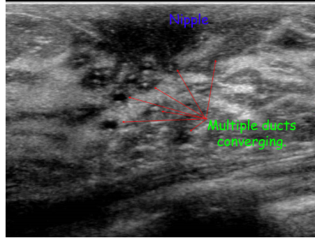
유두종 [ PAPILLOMA ]
섬유선종과 비슷한 경계가 분명한 결절인데, 대개 유륜 하부에 위치하며 늘어난 유관과 동반되어 보일 수 있습니다.
종괴 변연부에 석회화가 동반될 수 있습니다.
초음파상 늘어난 유관은 저에코의 관으로, 유두종은 고에코의 결절로 보입니다.
유관 내 병변을 찾기 위해 probe 를 유관의 주행을 따라 스캔해보아야 합니다.
Whist often benign, their malignant tendency generally leads to removal.
Multiple papillomas have been shown to carry a far greater risk than solitary.
–> Typically appear as a filling defect within a dilated duct or cyst


기타 유방염, 유방농양
유방염은 피부층의 두께 증가, 조직 경계면의 소실, 종괴를 형성하지 않은 길쭉한 저에코 소견 등이 보입니다.
유관확장증은 비화농성 염증 질환의 일종으로 유즙 분비나 통증의 원인이며,
양측 유관의 미만성 확장이 영상검사에서 보입니다.
농양이 형성되면 경계가 좀더 분명해지면서 유륜 하부에 저에코 종괴로 보이는데, 유방암과 구별이 어려울 수 있습니다.