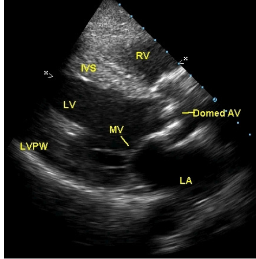
* Parasternal long axis view 에서 수축기에 doming 으로 관찰됨
Cusp calcification and thickening
Cusp motion limitation and doming
LVH
* Parasternal short axis view 에서 물고기 입 모양
Cusp calcification and thickening
Cusp motion limitation
Commissureal fusion (in Rheumatic AS)
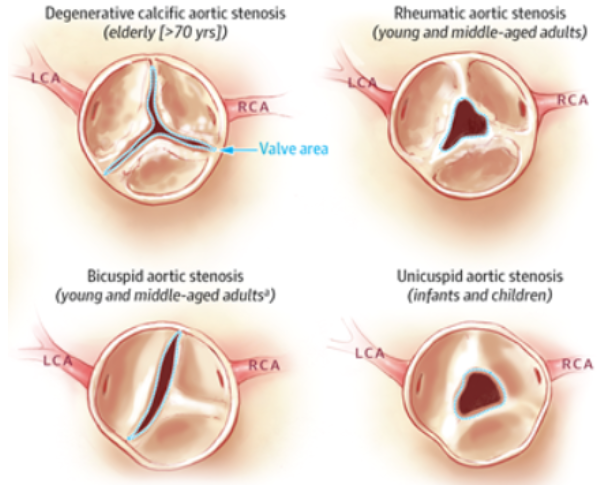
* Rheumatic AS 와 Degenerative AS 의 차이
Rheumatic AS 의 경우 commissural fusion, Multivalvular involvement
MV 를 더 먼저 침범하기 때문에 같이 involve 하는 경우가 많다.
촛농이 흘러내리는 모양
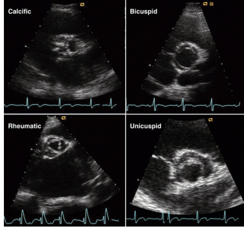
Parasternal mid-systolic short-axis view에서 calcific aortic stenosis는 leaflet의 aortic side에 발생한 fibro-calcific mass에 의해 특징지어지며, 이는 commissural fusion 없이 leaflet의 강직(stiffness)을 증가시킨다. Calcific shadowing과 reverberation으로 인해 영상의 질은 제한된다.
Congenital bicuspid valve의 경우, 두 개의 leaflet(앞쪽 leaflet에는 raphe 존재)이 수축기 동안 넓게 열리는 모습을 보인다.
Rheumatic stenosis의 진단적 특징은 commissural fusion과 mitral valve involvement이며, 수축기에서 특징적인 triangular aortic valve opening을 보인다.
Unicuspid valve는 단 하나의 부착 지점(6시 방향)만을 가지며, 깔때기 모양(funnel-shaped)의 판막 개구를 형성한다.
* Doppler Examination
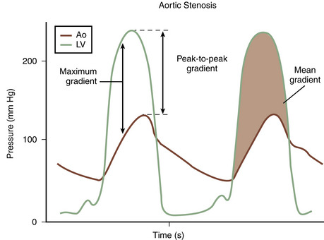
* CW doppler
– AS severity 평가시 중요
– 그림만 잘 그려주면 Peak pressure gradient, Mean pressure gradient 를 산출해 준다.
– Angle dependency 가 있으므로, 여러 방향에서 측정하여 도플러의 방향이 대동맥판을 지나는 혈류와 최대한 평행이 되도록 해야한다.
* LVOT obstruction 의 감별진단
– Valvular Aortic stenosis
– Subaortic membrane (Membrane 이나 web 이 생겨 obstruction)
– HCMP
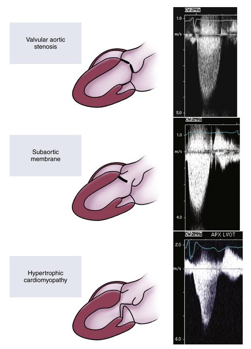
LV outflow obstruction의 다양한 유형
Valvular aortic stenosis, subaortic membrane으로 인한 fixed subvalvular obstruction, 그리고 hypertrophic cardiomyopathy로 인한 dynamic obstruction에서의 CW Doppler velocity curve 형태 예시이다.
Subvalvular obstruction과 valvular aortic stenosis의 CW 곡선은 유사하나, subvalvular obstruction에서는 판막의 coarse fluttering이 동반되어 수축기 속도 곡선이 “거친(rough)” 양상을 보인다. 이러한 감별은 2D 및 color flow imaging으로 가능하다.
Dynamic obstruction에서의 곡선 형태는 뚜렷하게 달라, 속도가 late systole에서 정점(peak)에 도달하는 특징을 보인다.
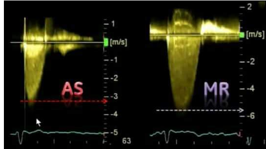
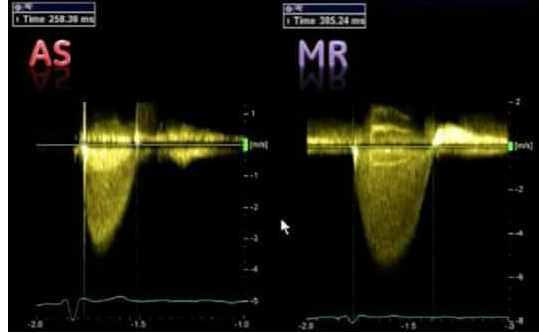
* High velocity systolic jets that may be mistaken for AS
– MR, TR, Supravalvular stenosis, VSD, Pulmonary artery stenosis, Peripheral vascular stenosis (e.g. Subclavian artery)
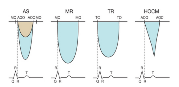
* AS jet vs. MR jet
AS jet : short duration, QRS 이후에 jet 이 시작
MR jet : wide, earlier, 보통 Vmax 가 5m/s 이상, QRS 바로 직후 혹은 동시에 jet 이 시작